FIG 3.
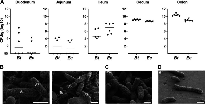
E. coli and B. thetaiotaomicron (Bt) colocalize in the mouse intestinal tract. (A) Bacterial loads in intestinal compartments. Intestinal samples were recovered from E. coli WT (Ec)- and B. thetaiotaomicron-cocolonized mice used in the experiment shown in Fig. 2, 72 h after the start of experiments. Intestinal contents were recovered from the five indicated locations of dissected mice, and CFU were determined. Bars represent the median values of CFU obtained from individual samples. ND, below the detection level. (B) Visualization by field emission scanning electron microscopy of feces from cocolonized mice. E. coli and B. thetaiotaomicron are identified by their distinct morphologies. Small particles may correspond to food particles or shed mucus. (C and D) Purified cultures were used for identification. White bars, 1 μM.

