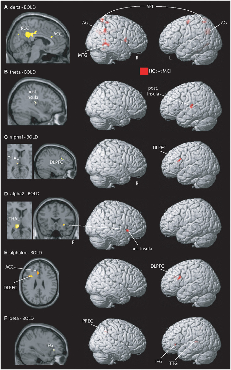
Figure 1.
Illustration of MCI-related fMRI signal coupling differences. Rows (A–D,F) shows results related to global spectral power (GSP) EEG-fMRI signal coupling; row (E) shows the local and band specific EEG-fMRI signal coupling. (A) Delta-fMRI signal alterations were seen in the posterior cingulate cortex (PCC), anterior cingulate cortex (ACC), precuneus (PREC), angular gyrus (AG), right frontal operculum, left pallidum, middle temporal gyrus (MTG), left lingual gyrus, fusiform gyrus, bilateral superior parietal lobe (SPL), superior occipital gyrus, precentral gyrus, and bilateral cerebellum exterior. (B) In theta, one cluster within the posterior insula (post. Insula) exhibited showed a coupling difference. (C,D) Alpha1 and alpha2 revealed altered coupling of the left dorsolateral prefrontal cortex (DLPFC), thalamus (THAL), right fusiform gyrus, and putamen. (E) Local alpha-BOLD signal was altered in the left DLPFC and ACC. (F) Beta EEG-fMRI signal correlations different in the left frontal operculum covering the inferior frontal gyrus (IFG) and anterior insular cortex (ant. Insula), transverse temporal gyrus (TTG), planum temporale, superior parietal lobe, and precuneus (PREC). For a detailed summary of all coupling results, see Table 2. Results are presented at p < 0.05 (cluster-corrected).

