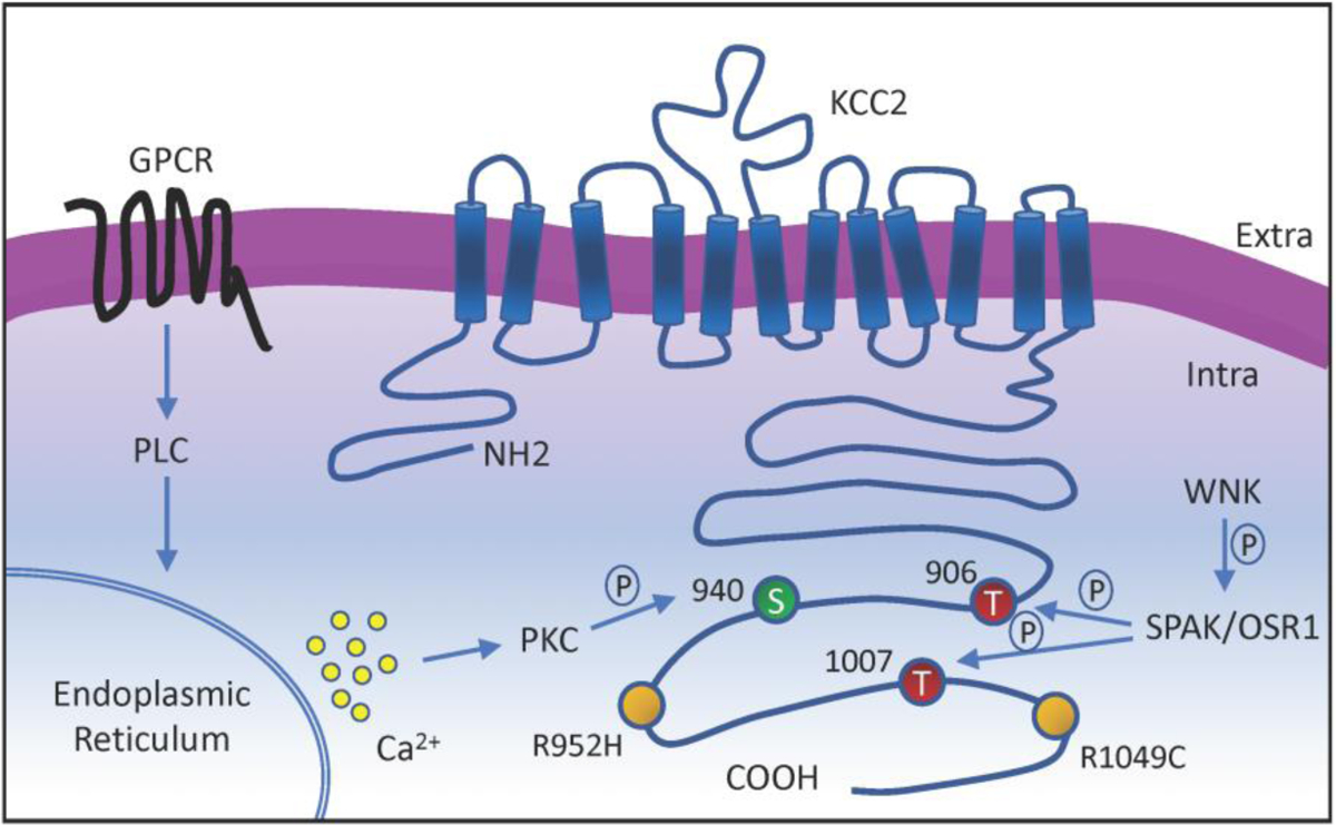
Figure 2: Membrane topology diagram of KCC2, potential therapeutic phosphorylation residues, regulatory pathways, and loss of function mutations associated with epilepsy.

The KCC2 co-transporter is a ~140 kDa protein with 12 transmembrane domains, a cytoplasmic N-terminal, a large extracellular loop, and a large cytoplasmic C-terminal. Phosphorylation residues regulated by WNK-SPAK pathway, which suppresses KCC2 activity, are indicated in red. Phosphorylation site regulated by neurotransmitters-GPCRs-PLC-PKC pathway, which increases KCC2 activity, is indicated in green. Missense variants of SLC12A5 that impair KCC2 function in patients with idiopathic generalized epilepsy are indicated in yellow.
