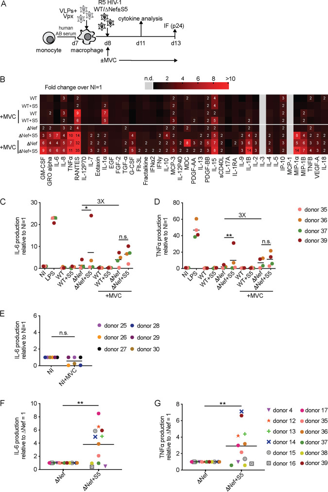
FIG 8.
Preventing fusion of HIV-1 ΔNef particles promotes proinflammatory cytokine production by MDMs. (A) Schematic of experimental flow (as in Fig. 5A, but in the presence or absence of MVC). (B) Cytokine expression in MDMs from donors 35, 36, 37, and 39 as a heat map. The color gradient covers up to a >10-fold change in expression over noninfected (NI; set to 1). Where indicated, cells were treated with MVC, starting from 1 h before infection for the entire experiment. (C and D) Quantification of IL-6 (C) and TNF-α (D) concentrations in the supernatant of infected MDM cultures relative to the NI control (set to 1). Shown are data points from cells of individual donors, with the mean of all donors indicated by a black line. The fold change between the ΔNef ± MVC conditions is indicated. (E) Concentration of IL-6 (relative to NI set to 1) in the supernatant of noninfected MDM cultures treated or not with MVC. Shown are data points from cells of individual donors, with the mean of all donors indicated by a black line. (F and G) Summary of the effects of S5 on proinflammatory cytokine production by HIV-1 ΔNef particles. Shown is the relative production of IL-6 (F) and TNF-α (G) for MDMs infected with HIV-1 ΔNef+S5 particles normalized to ΔNef (set to 1) from all 12 donors for which full cytokine profiles were established in this study. Data points indicate values from cells of individual donors, with the mean value for cells of all donors indicated by a black line. Statistics (Student's t test): n.s., nonsignificant; *, P < 0.05; **, P < 0.01.

