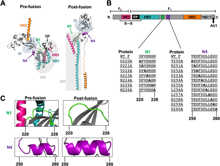
FIG 1.
Differences in spatial arrangements and secondary structures of the N1 and N4 regions between the pre- and postfusion conformations. (A) NiV F published prefusion and modeled postfusion monomer. The heptad repeats HR1 and -2 are highlighted in teal and orange, respectively. HR3 is highlighted in magenta. The N1 and N4 regions are highlighted in green and purple, respectively. The fusion peptide is shown in black. Areas shaded in gray denote the previously described DI and DIII domains (30, 31, 34). (B) Schematic of the NiV F protein. The green and purple areas denote the N1 and N4 regions, respectively. The N1 and N4 inserts show the native amino acid sequences as well as the scanning alanine mutants constructed in residues 220 to 226 and 250 to 260, respectively. WT, wild type. (C) Close-ups of spatial and/or secondary structural changes observed in the pre- and postfusion conformations of the N1 region in green and the N4 region in purple.

