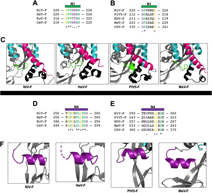
FIG 9.
N1 and N4 region sequence and structural homology. Sequence alignment of the portion of N1 in close association to the FP, HR1, and HR3 regions between NiV F and F of other henipaviruses (A) or between NiV F and F of other paramyxoviruses (B). (C) Structural positioning of the N1 region in the F prefusion structures among paramyxoviruses NiV, HeV, MeV, and PIV5, depicting the HR1 domain (teal), the FP domain (black), and the HR3 domain (magenta) (PDB accession codes 5EVM, 5EJB, 5YXW, and 4WSG). Sequence alignment of the portion of the N4 region between NiV F and F of other henipaviruses (D) or between NiV F and F of other paramyxoviruses (E). (F) Structural homology of the N4 region among paramyxoviruses in the prefusion conformation. The residues are either identical (*), very similar (:), or had similar residue side chains (.). PIV5 F was from the W3 strain, and MeV F was from the Ichinose-B95a strain. NDV, Newcastle disease virus; CDV, canine distemper virus.

