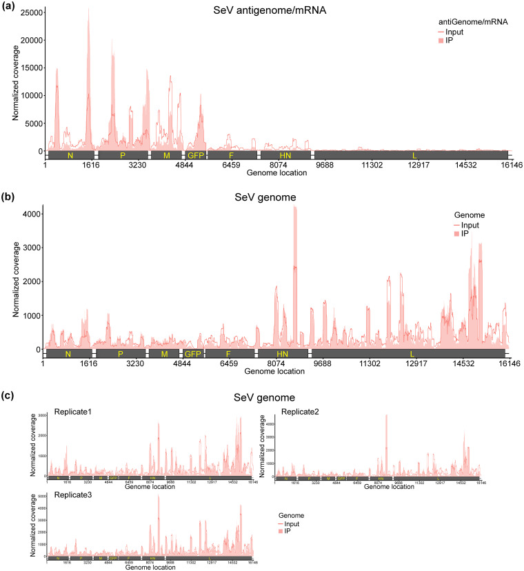
FIG 2.
The SeV RNAs are m6A methylated. (a and b), Distribution of m6A peaks in the SeV antigenome/mRNAs (a) and genome (b). A schematic diagram of the rSeV-GFP antigenome containing all genes (N, P, M, GFP, F, HN, and L) is shown. Total RNA was extracted from rSeV-GFP or mock-infected A549 cells and subjected to sonication. RNA containing m6A methylation was pulled down by m6A antibody, followed by m6A-seq. The shaded pink areas show the distribution of m6A immunoprecipitation (IP) reads mapped to the SeV antigenome/mRNAs (a) or genome (b). The baseline signal from input samples is shown as a line. Data presented in panels a and b are the averages from three independent virus-infected A549 cell samples (n = 3). (c) Distribution of m6A peaks in the SeV genome in individual replicates.

