
FIGURE 2.
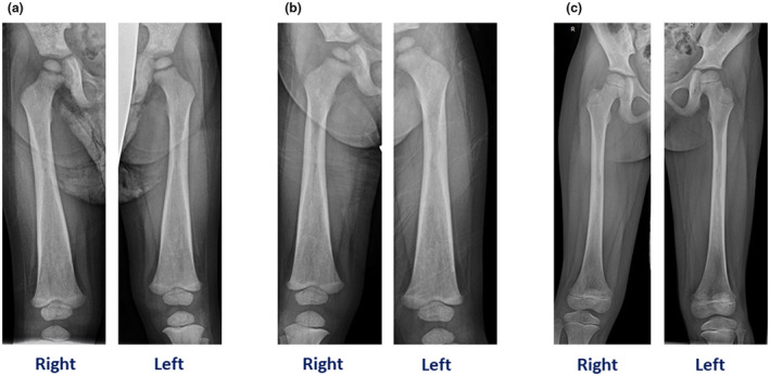
A, Patient 1. (a) skeletal survey revealed dense appearance of the long bones with diaphyseal and metaphyseal widening, particularly involving the femurs. (b) Patient 2. Dense appearance of the lone bones with diaphyseal and metaphyseal widening, especially involving femurs. (c). Patient 3. Patchy sclerosis and some cortical thinning involving the proximal to mid femoral diaphysis