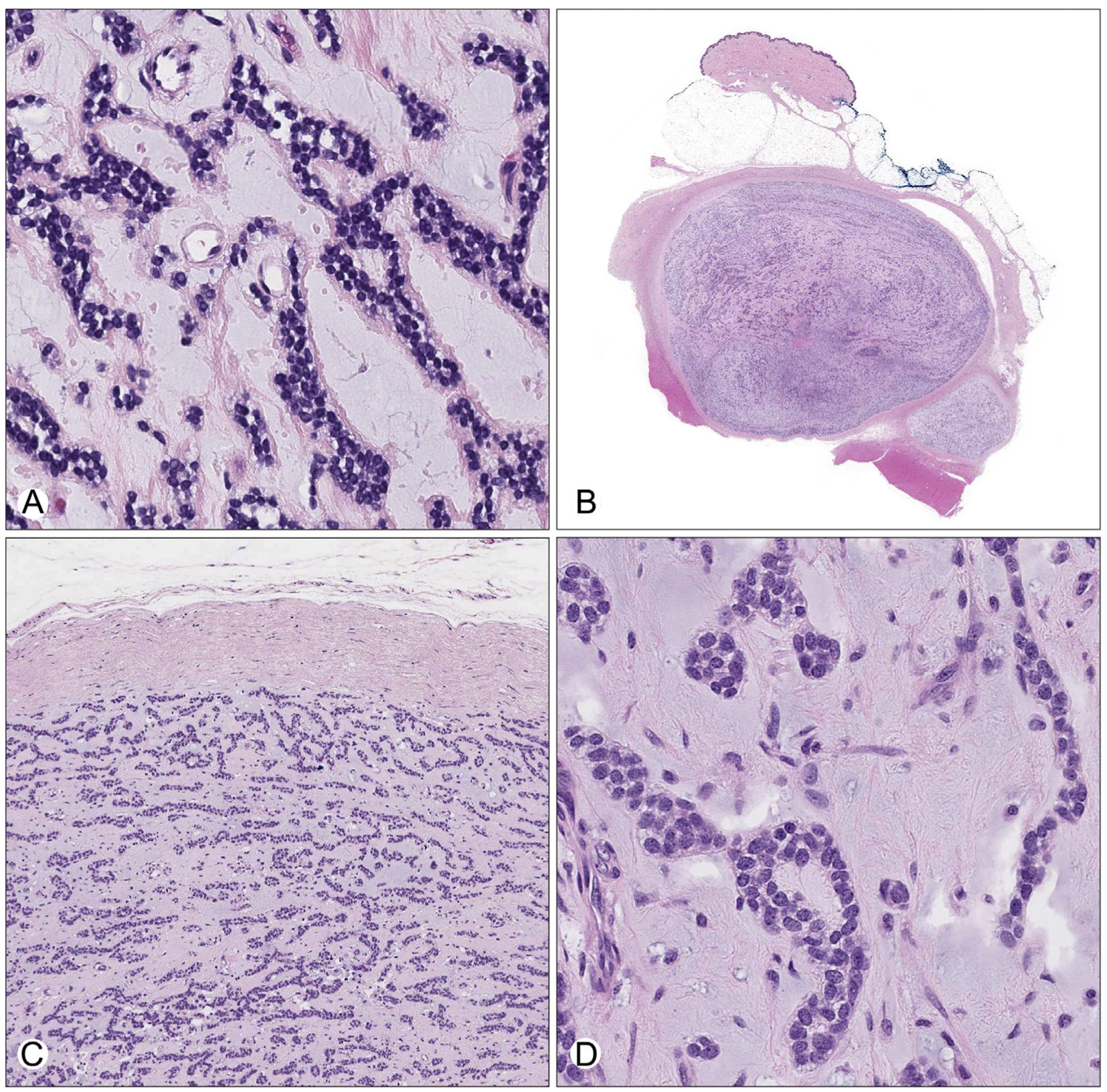
Fig. 2.

A, High-magnification view of the tumor in the incisional biopsy, characterized by anastomosing cords and pseudoacini of small round tumor cells embedded within a hypocellular myxoid stroma. B, Low-power view of the excision specimen showing a circumscribed, encapsulated subcutaneous mass with a pushing border into skeletal muscle. The modestly cellular tumor is embedded in a variable fibrous and myxoid stroma. C, Medium-power view demonstrating corded, interlacing thin-trabecular, and pseudoacinar growth patterns bounded by a thick fibrous capsule. D, High-power view showing monomorphic small round blue cells with fine, evenly dispersed chromatin, small nucleoli, and scant cytoplasm within abundant myxoid stroma.
