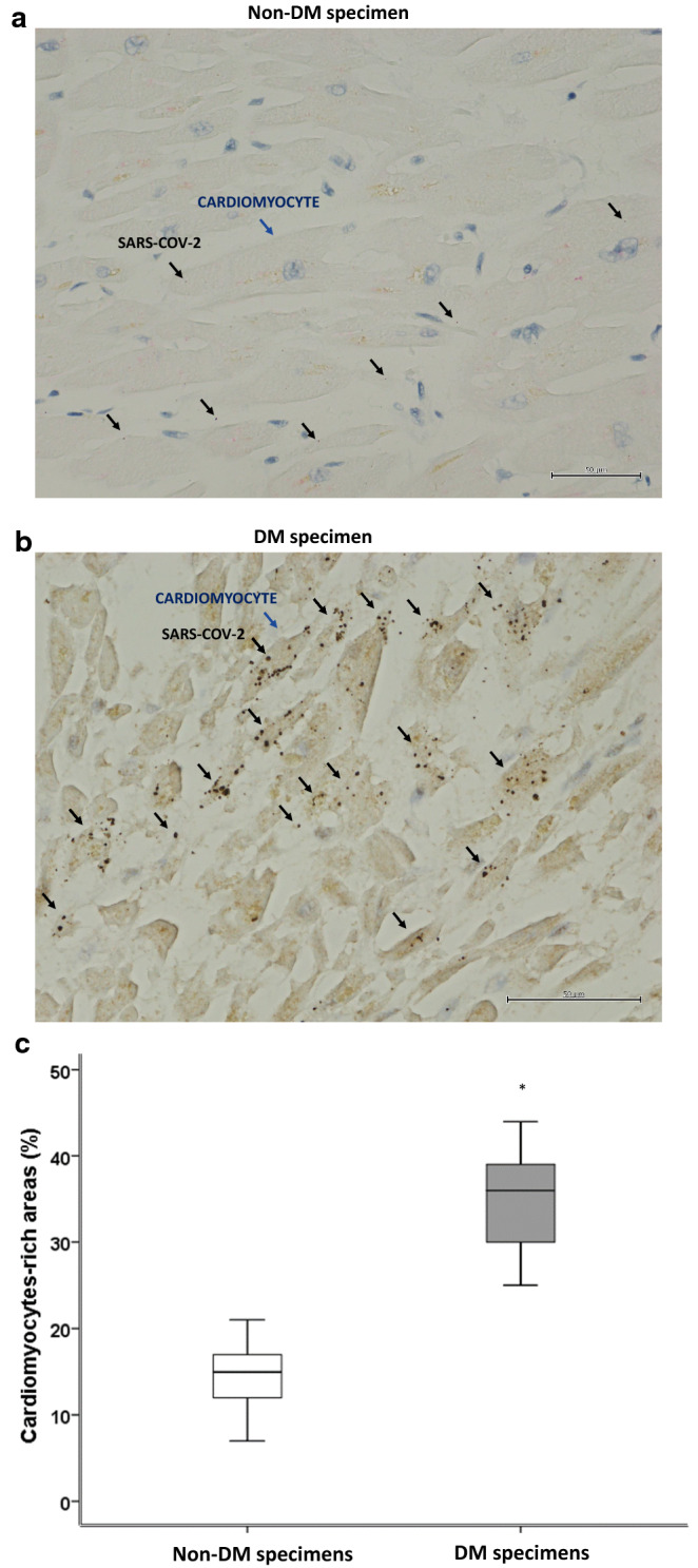
Fig. 1.

SAR-COV-2 in myocardial tissue from COVID-19 autopsies. a Representative myocardial tissue specimen from 60 patients without diabetes (Non-DM) (× 400). b Representative myocardial tissue specimens from 37 patients with diabetes (DM). Brown punctate evidenced the SARS-COV-2 RNA copies in the cardiomyocytes (96 positive cells/237 cells) (× 400). These structures are marked with black arrows (SARS-COV2), and with blue arrows (Cardiomyocytes). c Mean ± SD of the percentage of SARS-COV-2 positive cardiomyocyte. Statistical test: Student’s t-test. Bonferroni correction was used to make pairwise comparisons. *P < 0.05
