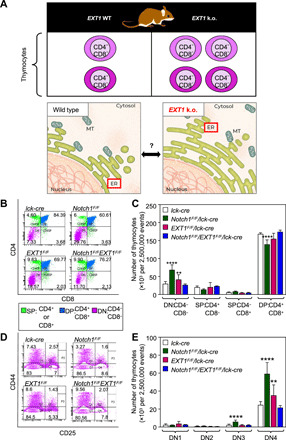
Fig. 1. Developmental defects following EXT1 inactivation are mediated by its genetic interactions.

(A) Framework to study the role of ER-resident EXT1 in thymocyte development. MT, mitochondria. (B) Representative fluorescence-activated cell sorting (FACS) plots showing the surface phenotype of CD4 and CD8 T cells in thymocytes. Cell percentages are shown in quadrants. (C) The absolute number of thymocytes (of 2,500,000 total events) showing the surface receptor expression of DN, single positive (SP) and double positive (DP) populations. n = 6 mice (lck-cre, Notch1F/F/lck-cre, EXT1F/F/lck-cre, and Notch1F/F/EXT1F/F/lck-cre). One-way ANOVA: **P < 0.01, ****P < 0.0001. (D) Representative FACS plots showing the surface expression of CD44 and CD25 markers in DN populations. Cell percentages are shown in quadrants. (E) The absolute number of DN1, DN2, DN3, and DN4 cells (of 2,500,000 total events). One-way ANOVA: **P < 0.01, ****P < 0.0001. See also fig. S1.
