
Fig. 4. DC-specific deficiency of c-Cbl promoted α-mannan–induced RelB activation to suppress IL-10 production for aggravating DSS-induced colitis.
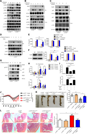
(A to B) Immunoblotting analysis of α-mannan–induced protein phosphorylation or nuclear translocation in BMDCs. (C) Immunoblotting analysis of α-mannan–induced nuclear translocation in DCs sorted from MLN in Cblf/fCD11cCre/+ and c-Cblf/f mice. (D) Immunoblotting analysis of α-mannan–induced protein phosphorylation or nuclear translocation in c-Cbl–deficient BMDCs treated with or without NF-κB–inducing kinase (NIK) inhibitor, isoquinoline-1,3(2H,4H)-dione. (E and F) ELISA results for IL-10 and IL-6 from BMDCs with indicated treatment (E) or knockdown with indicated small interfering RNAs (siRNAs) (F). (G) Immunoblotting analysis of α-mannan–induced interaction of p65 and RelB in BMDCs. (H) Chromatin immunoprecipitation assay of α-mannan–induced RelB and p65 binding to the promoter of il10 and il6 in BMDCs. (I) Luciferase assay of RelB- or p65-mediated activation of il10 or il6 promoters. (J to L) Weight loss (J), colon length (K), and histological analysis (L) of DSS (2.5%)–treated Cblf/fCD11cCre/+ and c-Cblf/f mice (n = 6 for each group), which were administrated with or without isoquinoline-1,3(2H,4H)-dione (50 mg/kg) at the indicated time. Bar graphs show means ± SEM. *P < 0.05, **P < 0.01, and ***P < 0.001. Photo credit: Jie-Lin Duan, Tongji University.