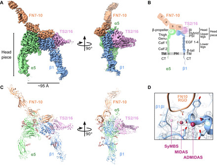
Fig. 1. Cryo-EM analysis of FN-bound integrin α5β1 reveals an open conformation.

(A) Two views of the FN7-10 (orange)–bound α5β1 (green, α subunit; blue, β subunit) cryo-EM structure, stabilized by TS2/16 (pink). The headpiece reveals an open conformation. (B) Schematic of integrin α5β1 domain architecture and nomenclature of different parts (headpiece, head, upper, and lower legs), including FN7-10 and TS2/16. TM, transmembrane; PM, plasma membrane; CT, cytoplasmic tail. (C) Molecular model based on the cryo-EM density, showing the same two views as (A). (D) Close-up and overlay of cryo-EM density with the modeled synergistic metal ion–binding site (SyMBS or LIMBS), MIDAS, and ADMIDAS within the βI domain.
