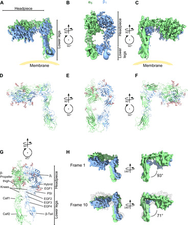
Fig. 4. Resting integrin α5β1 reveals an incompletely bent conformation.

(A to C) Three views of the resting integrin α5β1 cryo-EM structure. The position of the membrane is guided in yellow. Note that precise direction of the membrane cannot be determined because of the lack of membrane density from the structure. (D to F) The structure of the headpieces and modeled structure using homology models of PDB ID: 3fcs (13) for the lower legs. The headpiece reveals a closed conformation, with an orthogonal angle between the headpiece and legs. (G) The lower legs of integrin α5β1 are parallelly contacting each other, between EGF1/2 (β1) and calf1 (α5). (H) Multibody analysis revealed flexibility between the headpiece and lower legs, with an angle varying from 71° to 93° between the first analyzed state (“frame 1”) and the last state (“frame 10”). See fig. S7 for all the frames.
