Figure 4.
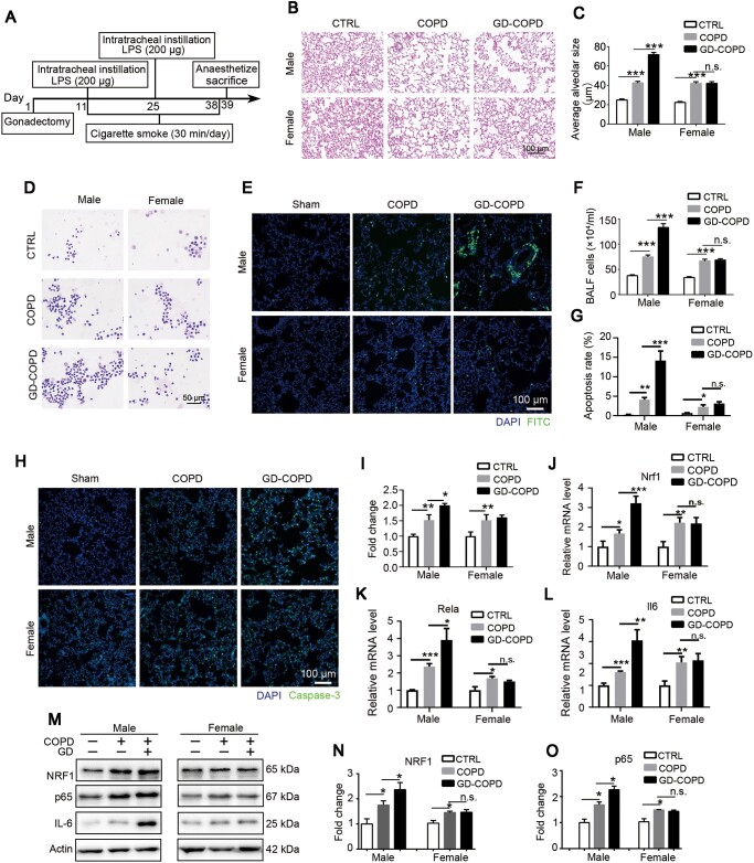
Absence of sex hormones exacerbates pulmonary lesions in male instead of female rats of COPD model. (A) Experimental design for the construction of castration in COPD model rats. (B and C) H&E staining was used to observe the morphological changes of COPD rats. Average alveolar size was measured and statistically analyzed (200 alveoli per animal were counted). (D and F) Wright staining was used to observe the change of cell number and leukocyte type in BALF. (E and G) Apoptotic cell nuclei were stained with FITC fluorescence by TUNEL assay. (H and I) Pulmonary tissues were stained with caspase-3 antibody and intensity of green signal was statistically analyzed. (J‒L) mRNA expression levels of Nrf1, Rela, and Il6 were measured by qRT-PCR. (M‒O) Protein levels of NRF1 and p65 were determined by western blotting. n = 6, mean ± SEM, n.s. means no significance, *P ≤ 0.05, **P ≤ 0.01, ***P ≤ 0.001 by one-way ANOVA.

