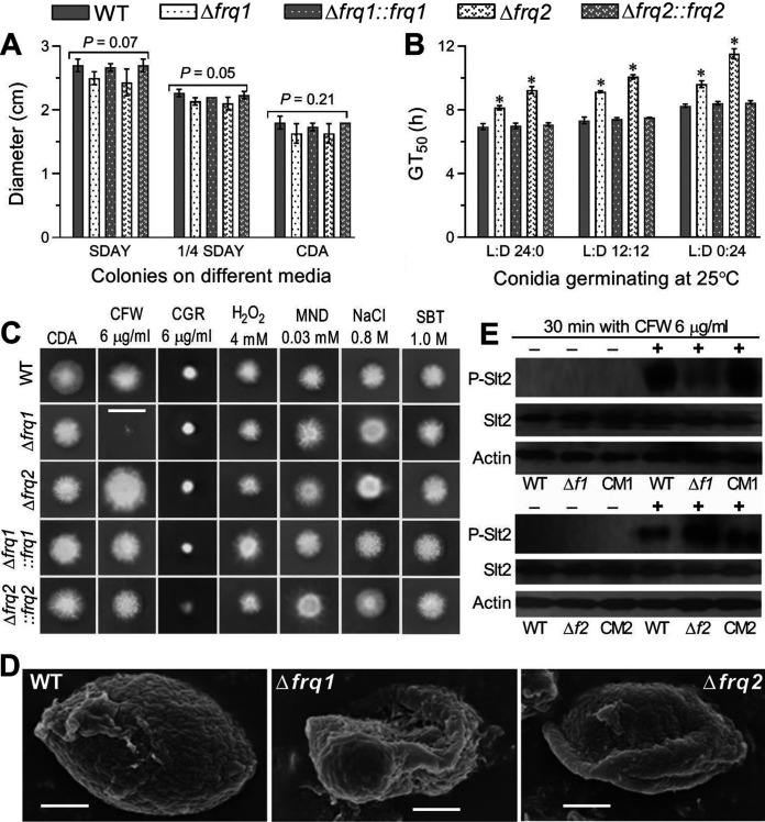
FIG 3.
Impacts of frq1 or frq2 deletion on the growth of B. bassiana under normal and stressful conditions. (A) Diameters of fungal colonies after an 8-day incubation on SDAY, 1/4 SDAY, and CDB at the optimal regime of 25°C and L:D 12:12. (B) Time for 50% conidial germination (GT50) at 25°C in the three indicated L:D cycles. (C) Images (scale bar, 2 cm) of fungal colonies grown at 25°C for 10 days on CDA containing CFW (calcofluor white), CGR (Congo red), H2O2, MND (menadione), NaCl, and SBT (sorbitol) at the indicated concentrations, respectively. Each colony was initiated by spotting 1 μl of a 106 conidia/ml suspension. (D) SEM images (scale bar, 0.5 μm) for ultrastructural views of conidial surfaces. Note the increased fragility and reduced plasticity of conidial coat for the Δfrq1 and Δfrq2 mutants, respectively, in comparison to an integrity of conidial coat for the WT strain. (E) Western blots of phosphorylated Slt2 (P-Slt2), expressed Slt2, and expressed β-actin (reference) in the hyphal cells triggered for 30 min with (+) or without (−) CFW (6 μg/ml). Each lane was uploaded with 60-μg protein extract. Δf1 and CM1, Δfrq1 and Δfrq1::frq1. Δf2 and CM2, Δfrq2 and Δfrq2::frq2. The means represented by asterisked bars in each bar group (B) differ significantly from those of the unmarked bars (Tukey’s HSD, P < 0.01). Each error bar (A, B) indicates standard deviation of the mean from three independent replicates.

