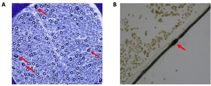
Figure 2.
Nerve biopsy from a CADM3 patient. (A) A cross-section of sural nerve from a patient of Family 2 showing a reduced number of large myelinated fibres and very few groups of small regenerating fibres. Of interest, we overserved a number of abnormally myelinated fibres with thick folded myelin. A teased fibre from the same biopsy (B) shows such a focal myelin swelling or hypermyelination, a histological feature of demyelinating peripheral neuropathies.

