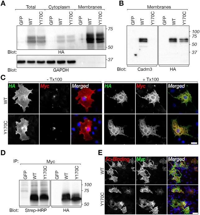
Figure 4.
CADM3 mutant reaches the cell surface of transfected cells and interacts with CADM4. (A) Western blot analysis of total cell lysate (Total), cytoplasmic and membrane preparation showing the expression of HA-tagged (tag located extracellularly) and Myc-tagged (tag located intracellularly) of rat CADM3 (wild-type, WT) and CADM3 mutant (Y170C) in transfected HEK293 cells, using an antibody to HA or to GAPDH as indicated. Cells transfected with green fluorescent protein (GFP) were used as control. Molecular size markers (kDa) are shown on the right. (B) Western blot analysis of membrane preparations from HEK293 cells expressing the same constructs as in A, using a polyclonal antibody directed to the extracellular domain of CADM3 or an antibody to HA-tag as indicated. Note that the CADM3 antibody recognize the wild-type but not the mutant CADM3 protein. Molecular size markers (kDa) are shown on the right. (C) Both wild-type and mutant proteins are present at the cell surface. Immunolabelling of HEK293 cells transfected with the double-tagged versions of wild-type or mutant CADM3 was carried out using either live (− Tx100) or permeabilized (+ Tx100) cells. The single channels and merged images are shown as indicated. (D) Surface biotinylation of HEK293 cells transfected with wild-type (WT), mutant (Y170C), or GFP, followed by immunoprecipitation (IP) with an antibody to Myc and immunoblot using streptavidin-HRP (Strep-HRP) or an anti-HA antibody as indicated. Molecular size markers (kDa) are shown on the right. (E) The mutant CADM3 interacts with the extracellular domain of CADM4. COS7 cells expressing Myc-tagged wild-type (WT) or mutant (Y170C) CADM3 were incubated with medium containing a soluble extracellular domain of CADM4 fused to the Fc region of human IgG. Expression of CADM3 and binding of Cadm4-Fc was monitored using antibodies to human Fc (Fc-Binding) and Myc tag, as indicated. DAPI labelling (blue) of cell nuclei is shown in the merged images. Scale bars = 20 μm.

