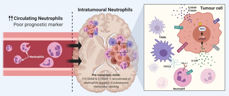
Figure 3.
Role of neutrophils in brain metastases. Left: In the setting of brain metastases, a high neutrophil-to-lymphocyte ratio is associated with poor prognostic outcomes. Middle: In the tumour microenvironment, neutrophils are recruited through increased release of S100A8 and S100A9 proteins by tumour cells. Right: The increased expression of CD117, ADORA2A, ITGA3 and MET by brain metastatic cells leads to neutrophilic suppression in the immune microenvironment. The phosphorylation of EZH2 protein increases the expression of c-JUN, which leads to the release of G-CSF. G-CSF, in turn, leads to the recruitment of neutrophils that overexpress PD-L1 and Arg1. Furthermore, tumour-associated myeloid cells and microglia release chemokines, such as CXCL8, to suppress neutrophils in the tumour microenvironment. Created with BioRender.com.

