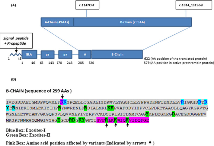
FIGURE 2.

Structure of prothrombin protein and effect of variants on protein. (A) This figure shows the domain architecture of the prothrombin protein with location of the two variants (identified in our patient) in the B‐chain region of the protein. (B) Amino acid sequence of the B chain with highlighted (blue and green) exosite‐I and II positions and pink boxes highlighting the exosite‐I/II positions affected by these two variants
