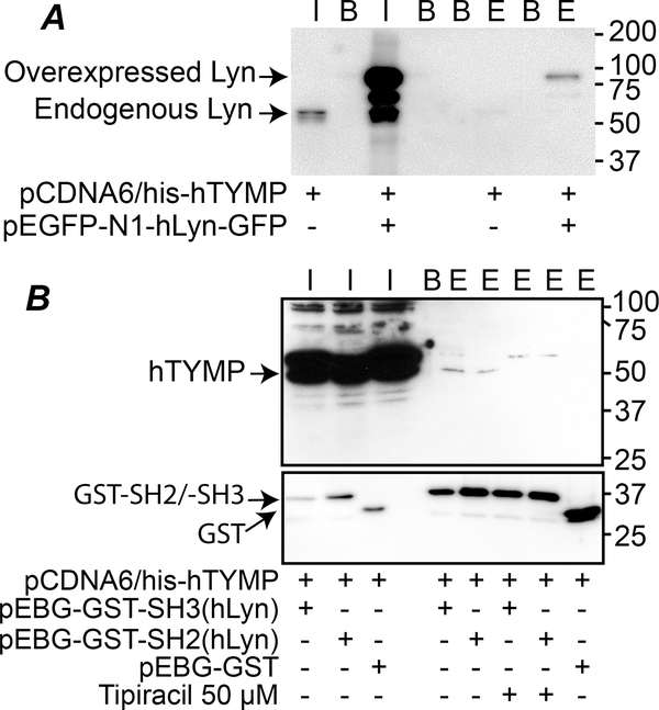
Fig. 1. TYMP binds to its partner through their SH3 or SH2 domain.
A. pCDNA6/his-hTYMP plasmid vector, either alone or combined with pEGFP-N1-hLyn-GFP vector, was transfected into Cos-7 cells, and His-Tagged TYMP was pulled down using His Mag Sepharose Ni beads. Inputs and elutes were blotted using anti-Lyn antibody. B. pEBG-GST-SH3(hLyn), pEBG-GST-SH2(hLyn), or pEBG-GST empty vector were co-transfected with pCDNA6/his-hTYMP into Cos-7 cells with or without TPI (50 μM) in culture media. The cell lysates were used for GST pull-down assays and eluted TYMP was detected by western blot using an anti-human TYMP antibody. In both panels, I: input; B: blank (2x Laemmli sample buffer only), E: elute. Blots represents 2–3 repeats.

