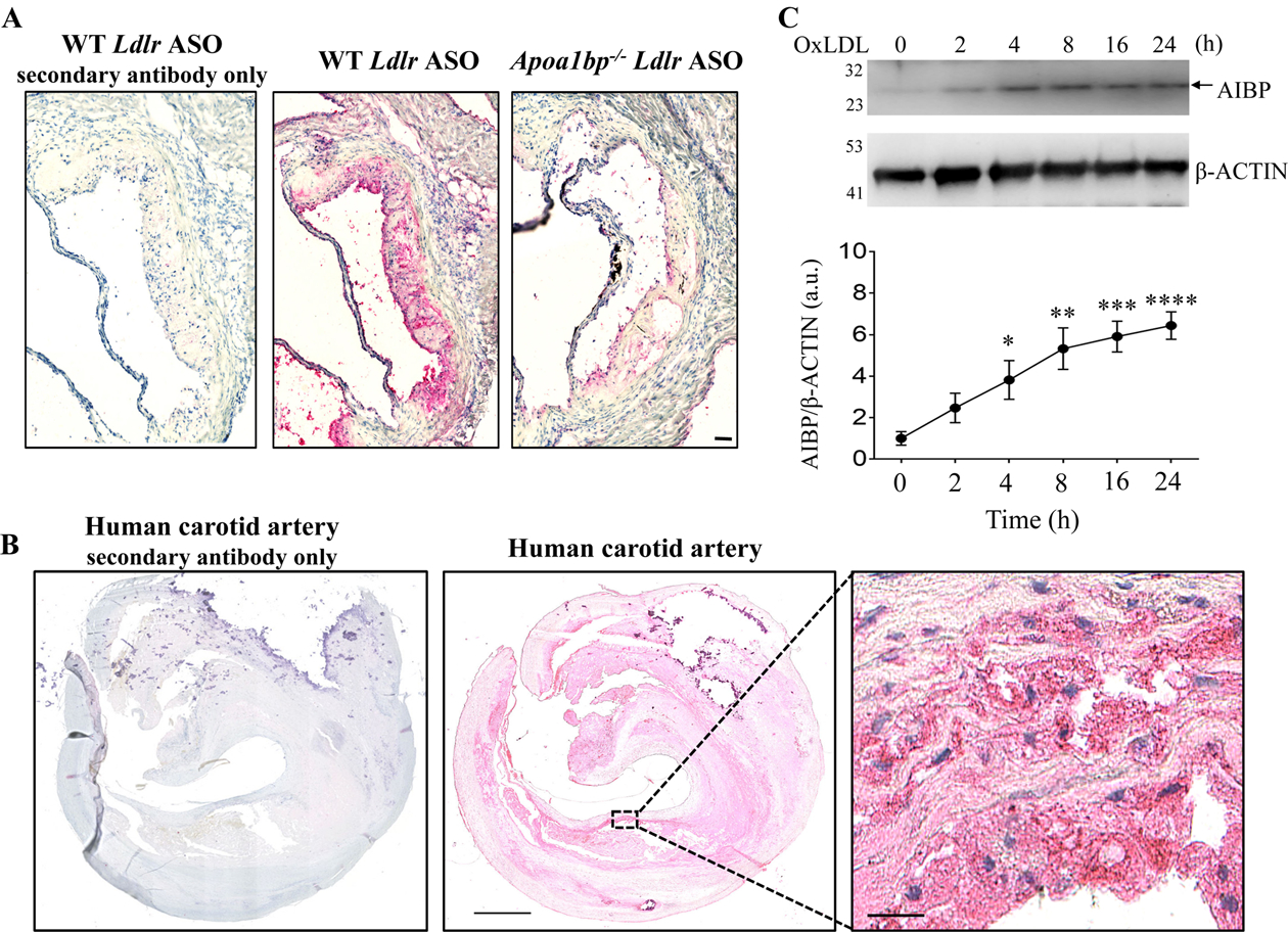
Figure 1.

AIBP expression in atherosclerotic lesions and in OxLDL-stimulated macrophages. Sections of the aortic root of hypercholesterolemic WT and Apoa1bp−/− mice (A) and human carotid artery (B) were stained with the monoclonal anti-AIBP antibody BE-1, followed by a secondary antibody or by the secondary antibody only, and counterstained with H&E. Representative images of three specimens tested. (C) BMDM isolated from WT mice were stimulated with 25 μg/ml OxLDL for indicated time. Cells lysates were immunoblotted with anti-AIBP and β-ACTIN antibodies. Band intensities were quantified. Mean±SEM; N=4–5. *, p<0.05; **, p<0.005; ***, p<0.0005; ****, p<0.0001 vs. time zero. Scale bars: 50 μm in A, 1 mm in B, and 25 μm in the zoomed-in region in B.
