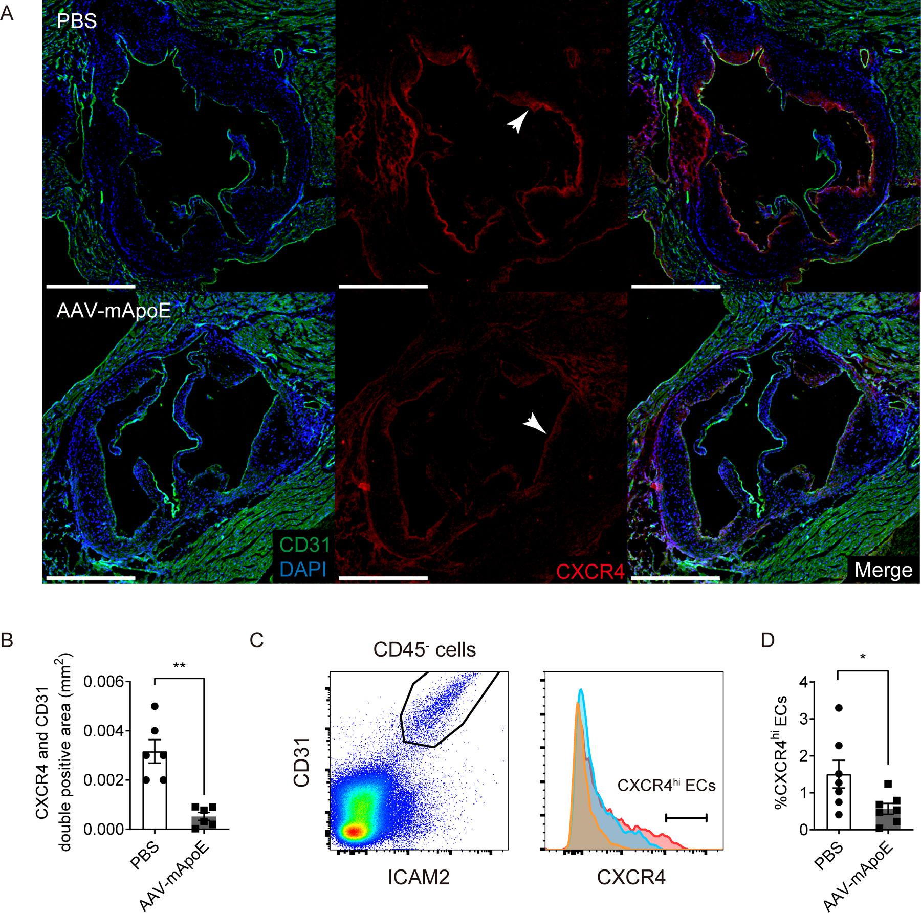
Figure 5. Plaque regression is associated with loss of CXCR4 on aortic endothelium.

(A) Representative images of CXCR4 expression in aortic sinus from Apoe−/− mice on HFD after 2 weeks of PBS (upper) or AAV-mApoE (bottom) treatment. Scale bar: 500 μm, and (B) quantification of CXCR4+ CD31+ areas (n=6 per group). (C) Representative histogram of CXCR4 expression on aortic ECs from Apoe−/− mice on HFD after 2 weeks of PBS (red) or AAV-mApoE (blue) treatment. Yellow histogram is isotype control. (D) The percentage of CXCR4hi ECs in total aortic ECs with or without 2 weeks of AAV-mApoE treatment (n=7 per group). All data are shown as means ± SEM. *p<0.05, **p<0.01 by Mann-Whitney U test.
