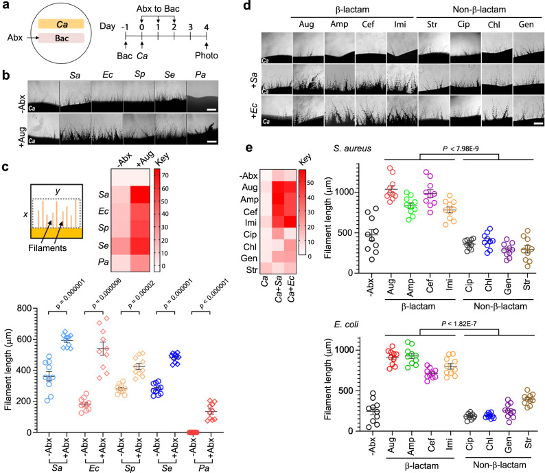
Fig. 1. Bacteria promote C. albicans hyphal growth and the activity is significantly enhanced by β-lactam antibiotics on plates.
a Experimental design. 1 × 10⁶ bacterial cells were inoculated into a 0.5 cm × 4 cm area YPD plate and grown at 37 °C for 24 h. Then, C. albicans yeast cells were streaked alongside the bacterial patch for incubation at 30 °C for 4 d. See Supplementary Table 1 for all C. albicans and bacterial strains used in this study. b Microscopic images of C. albicans filaments on day 4. Scale bar, 250 µm. The experiment was repeated independently three times with similar results. c Quantification of C. albicans filamentation shown in (b). First, a fixed area of all images was scanned using the mean gray area function in ImageJ. x was determined by the average length of ten randomly selected filaments in the image with the strongest filamentous growth, and y is the width of the image. The intensity values of all images were normalized against that of C. albicans without antibiotic (Abx) treatment and shown by the heatmap (see Methods for detail). Second, 10 filaments (n = 10) in each image were randomly chosen to measure the length. P values were calculated using two-tailed unpaired t test. Error bars = means ± SEM. d C. albicans was grown alone or side-by-side with Sa or Ec with or without antibiotic treatment. The results were analyzed as described in (a). The antibiotic solution (250 µg for all antibiotics used) was dropped evenly onto the surface of the bacterial patch at 24 h of growth followed by inoculating C. albicans. Scale bar, 250 µm. e Quantification of filamentous growth as shown in (c). Ten filaments (n = 10) were measured for each treatment. P values were calculated using two-tailed unpaired t test. Error bars = means ± SEM.Source data are provided as a Source Data file.

