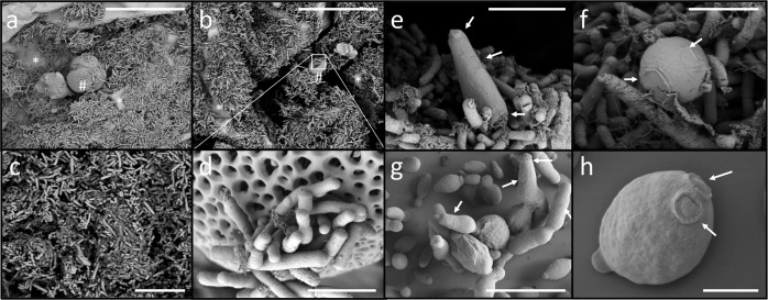
Fig. 1. Visualization of microorganisms inhabiting honeybee guts.
a–d Scanning electron microscope (SEM) micrographs showing bacterial cells present in a honeybee gut (i.e., rod-shape bacteria). Grains of pollen covered by bacteria are shown (see also Supplementary Fig. S1). e, f Fungal cells (i.e., yeast) associated with the gut wall of a honeybee. g, h Culture of yeast cells isolated from the honeybee gut. Cells belong to Hanseniaspora uvarum (strain L18) and Starmerella bombicola (strain L28; see also Supplementary Fig. S9 and Supplementary Table S7). Note the different scales on the SEM photographs. Honeybees used for SEM analysis were A. mellifera jemenitica from Saudi Arabia (Supplementary Table S6). Scale bars correspond to (a, b) 30 µm, (c) 3 µm, (d, f) 2 µm, (e, g) 10 µm, and (h) 1 µm.

