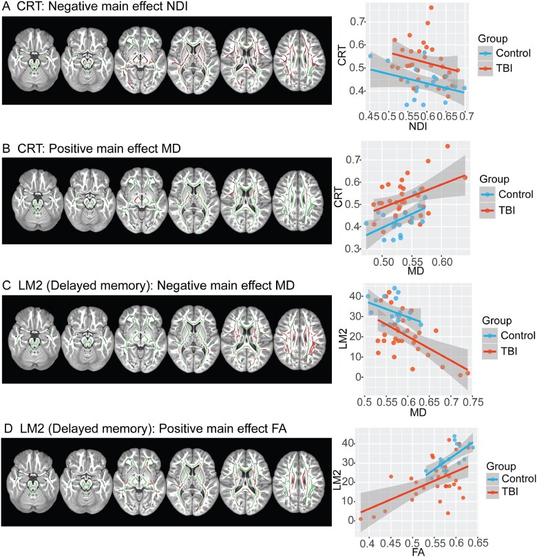
Figure 6.
Correlation of neuropsychological assessment with MRI measures of WM. (A) Voxels with a negative correlation between NDI and CRT (red); (B) Voxels with a positive correlation between mean diffusivity and CRT; (C) Voxels with a negative correlation with delayed recall logical memory; (D) Voxels with a positive correlation and delayed recall on logical memory. Contrasts are overlaid on the mean FA skeleton (green) and are adjusted for age, gender and intracranial volume (TFCE: P < 0.05, corrected for multiple comparisons). Scatterplots illustrate the mean intensity values of significant voxels against cognitive performance for each of the tests.

