Figure 3.
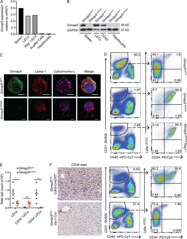
Genetic deficiency of GIMAP5 causes liver endothelial cell abnormalities. (A) Gimap5 mRNA expression in sorted liver endothelial cells (LECs; DAPI−CD45−CD31+) from C57BL/6 WT and Rag1−/− mice, and splenocytes, sorted Kupffer cells (DAPI−CD45+CD115+F4/80+), and hepatocytes from C57BL/6 mice. (B) Immunoblot for GIMAP5 in Gimap5sph/+ and Gimap5sph/sph splenocytes and hepatocytes and in sorted LECs and liver CD45+ cells from Gimap5sph/+ mice. GAPDH is shown as a loading control. (C) Confocal microscopy of LECs isolated from Gimap5sph/+ and Gimap5sph/sph and stained for GIMAP5 (green), Lamp-1 (red), and cytochrome-c (magenta) and counterstained with DAPI (blue). Scale bars = 5 µm. (D) LECs isolated from Gimap5sph/+, Gimap5sph/sph, and Gimap5sph/sphRag1−/− livers (left panels) and respective Ly6a and CD34 surface expression (right panels). (E) Absolute number of LECs that express CD34 or not in Gimap5sph/+ (n = 6) and Gimap5sph/sph (n = 5) livers. (F) Histological and flow cytometric analysis of sinusoidal CD34 expression in tamoxifen-treated adult Gimap5flx/flxxCdh5(PAC)-CreERT2 and Gimap5flx/flxxVav1-Cre mice (upper and bottom panels, respectively). Experimental data were verified in at least two independent experiments, and littermates were used as controls. Scale bars = 50 µm. Numbers depict the percentage of total cells. Student’s two-tailed t test was used. *, P < 0.05; **, P < 0.005; ***, P < 0.0005. Adult mice, >7 wk old. APC, allophycocyanin; Cy, cyanine.

