Figure S2.
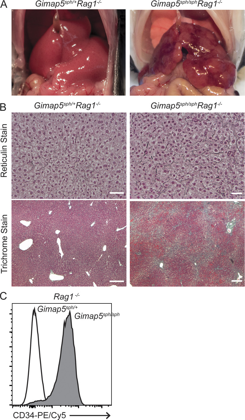
Liver pathology of Gimap5sph/+Rag1−/− and Gimap5sph/sphRag1−/− mice. (A) Gross liver morphology of adult Gimap5sph/+Rag1−/− (smooth) and Gimap5sph/sphRag1−/− mice (nodular). (B) Reticulin and trichrome stains of liver sections from Gimap5sph/+Rag1−/− (left panels) and Gimap5sph/sphRag1−/− (right panels) show two-cell-thick plate and increased collagen (blue) deposition solely in mutant mice. (C) Flow cytometric analysis shows increased CD34 expression in liver endothelial cells (DAPI−CD45−CD31+) isolated from Gimap5sph/sphRag1−/− mice as compared with Gimap5sph/+Rag1−/− mice. Experimental data were verified in at least two independent experiments, and littermates were used as controls. Scale bars = 50 µm.

