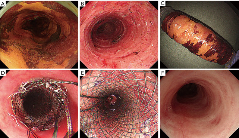
Figure 2.
One case of prevention of ES was conducted with PGA AEM transplantation and TSI. (A) Lugol iodine staining showed a complete annular lesion in the esophageal lumen. (B,C) ESD caused a wholly circumferential mucosal defect. (D) The stent was placed at the site of the artificial esophageal ulcer. (E) The patient underwent a weekly endoscopy to confirm that the stent was in place. (F) No evidence of stenosis was observed 6 months after transplantation.

