
Fig. 6 ∣. OP-Puro incorporation by HSCs and myeloid progenitor cells in vivo.
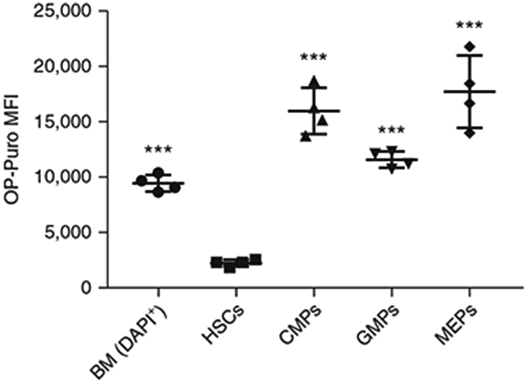
MFI of OP-Puro in unfractionated bone marrow cells (BM), HSCs, CMPs, GMPs and MEPs (n = 4). Background fluorescence has been subtracted from each population. The raw data are shown in Tables 5 and 6. Data represent mean ± standard deviation. Statistical significance relative to HSCs was assessed using a one-way ANOVA followed by Dunnett’s test for multiple comparisons; ***P < 0.001. All procedures in this protocol involving mice were approved by the UC San Diego Institutional Animal Care and Use Committee.