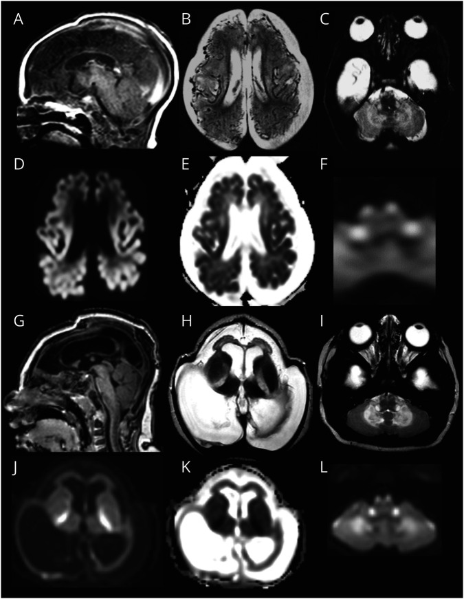
Figure 1. Prototype MRI for Group 1.
In patient LBSL285 at age 2 days, the cerebrum is hypoplastic (A and B). Numerous abnormal, tortuous blood vessels are seen at the surface (B). The cortex is hardly discernible from the white matter (B). Diffusion restriction is present in the cerebral cortex (diffusion-weighted image in D, ADC map in E). Tracts in the brainstem are affected; shown are the pyramids and inferior cerebellar peduncles (C), which also display diffusion restriction (F). On follow-up at 5 months, the cerebral mantle is reduced to a thin rim (G and H). The abnormal vessels are no longer visible (H). The pyramids and inferior cerebellar peduncles are still abnormal (I). Diffusion restriction is shown in the posterior limb of the internal capsule, cerebellar white matter, and brain stem tracts (diffusion-weighted images in J and L; ADC map in K).

