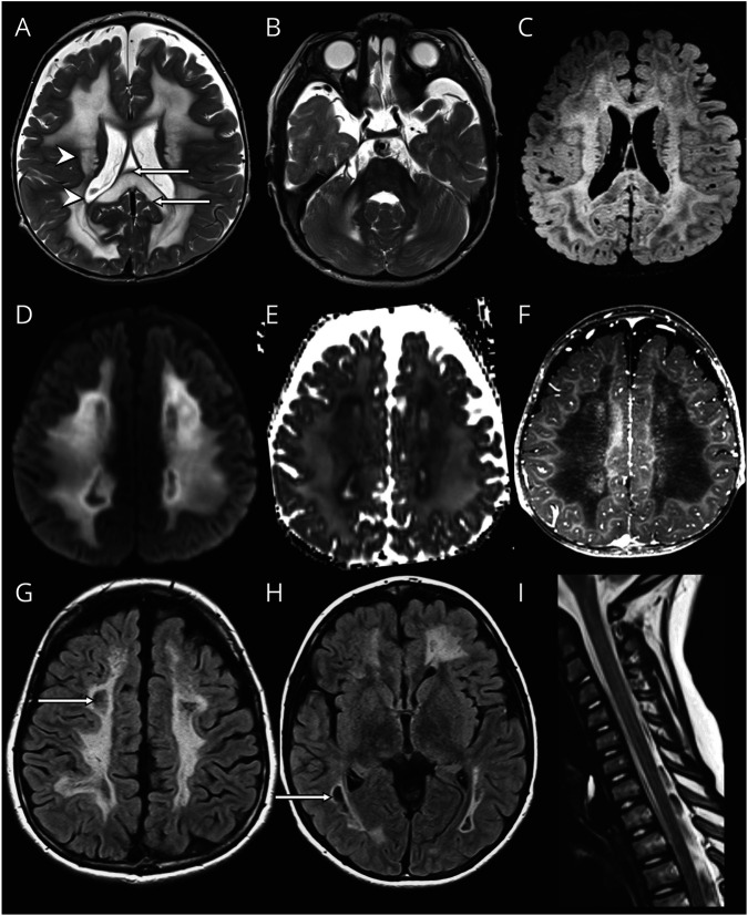
Figure 2. Prototype MRI for Group 2.
In patient LBSL288 at age 1 year and 2 months, the cerebral white matter is rarefied but not cystic (FLAIR image in C). The directly periventricular and directly subcortical rims are unaffected (arrowheads in A), and the middle blade of the corpus callosum is affected, whereas the inner and outer blades are spared (arrows in A). There is no brainstem involvement (B). The area directly adjacent to the rarefied white matter shows restricted diffusion (diffusion-weighted image in D, ADC map in E) and enhancement after contrast (F). On follow-up MRI at 8 years, rarefaction and cystic degeneration in white matter is present (arrows in FLAIR images in G and H). There is no spinal cord involvement (I).

