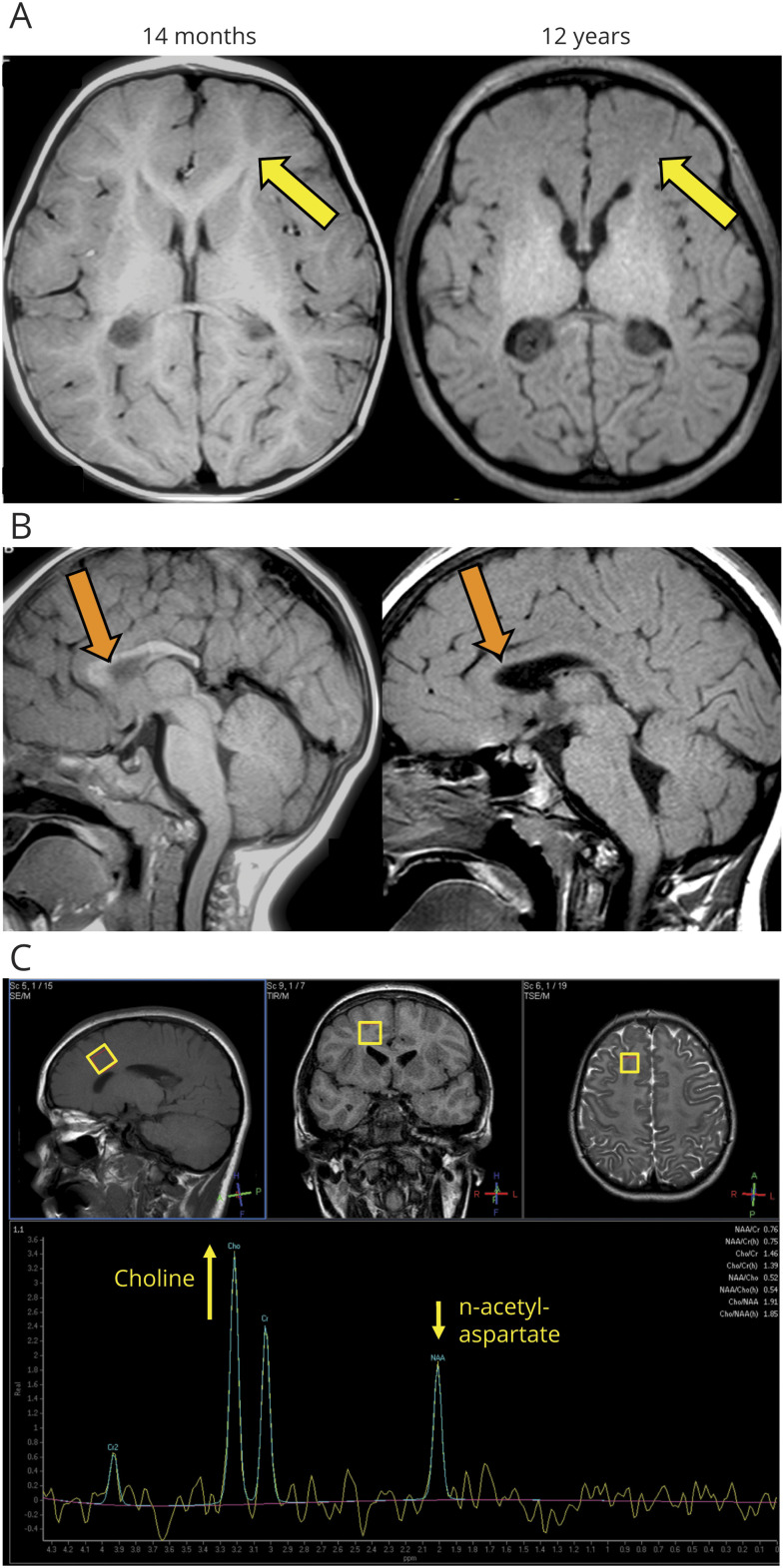
Figure 3. Brain MRI of Patient 1 Shows Corpus Callosum Dysgenesis and Loss of Supratentorial Myelin.
(A) T1-weighted MRI showing normal myelination at 14 months but loss of normal T1 hyperintense white matter signal at 12 years consistent with severe hypomyelination (yellow arrows). (B) T1-weighted MRI showing dysgenesis of the corpus callosum with a deficient splenium. Although there is normal myelin in the genu of the corpus callosum at 14 months, it is lost at 12 years (orange arrows). In addition, there is platybasia with resultant kinking of the medulla oblongata. (C) Single voxel proton MR spectroscopy with TE 144 ms at 12 years of age. Top row: T1W, T2 FLAIR, and T2W images show abnormal white matter. Bottom row: spectrum shows elevated choline relative to n-acetylaspartate (NAA) in the right frontal lobe white matter, consistent with the loss of sphingolipid in the myelin and suggestive of myelin breakdown. A normal spectrum should show reversed levels of NAA and choline.17.

