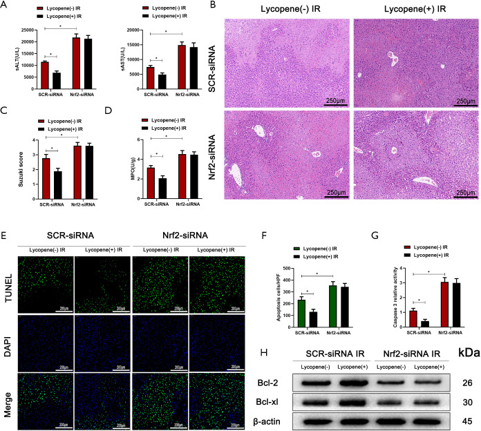
Figure 6.
Nrf2 knockdown in Kupffer cells (KCs) exacerbates IR-induced acute liver injury. (A) Serum ALT and AST in mice who underwent IR injury in different groups (n=5 mice/group). (B) Representative haematoxylin and eosin (HE) staining of liver tissue sections from different groups (scale bars, 250 µm). (C,D) The average Suzuki scores and liver myeloperoxidase (MPO) activities of different groups. (E,F) TUNEL staining of liver sections (scale bars, 200 µm) and the relative ratios of TUNEL-positive cells in different groups. (G) Relative caspase-3 activity was evaluated from total lysates of IR lobes from different groups. (H) Western blot analysis of the protein levels of Bcl-2, Bcl-xL, and β-actin in different groups. All data represent the mean ± SD, *P<0.05. IR, ischemia reperfusion; ALT, alanine aminotransferase; AST, aspartate aminotransferases.

