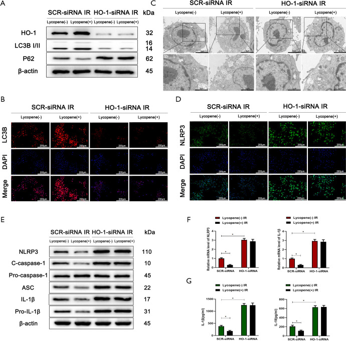
Figure 7.
HO-1 knockdown inhibits Nrf2-mediated autophagy and promotes the expression of the NLRP3 inflammasome in Kupffer cells (KCs). (A) Western blot analysis of the protein levels of HO-1, LC3B, p62, and β-actin in KCs isolated from mice who underwent IR injury in different groups (n=5 mice/group). (B) Immunofluorescence staining of LC3B in KCs isolated from different groups (scale bars, 200 µm). (C) Autophagic microstructures in KCs isolated from different groups detected by transmission electron microscopy (scale bars, 5 and 2 µm). (D) Immunofluorescence staining of NLRP3 in KCs isolated from different groups (scale bars, 200 µm). (E) Western blot analysis of the protein levels of NLRP3, cleaved caspase-1, procaspase-1, ASC, IL-1β, pro-IL-1β, and β-actin. (F) Quantitative RT-PCR analysis of the relative mRNA levels of NLRP3 and IL-1β in KCs isolated from different groups. (G) Levels of IL-1β and IL-18 in the culture supernatants of KCs isolated from different groups after 6 h of culture measured by ELISA. All data represent the mean ± SD, *P<0.05.

