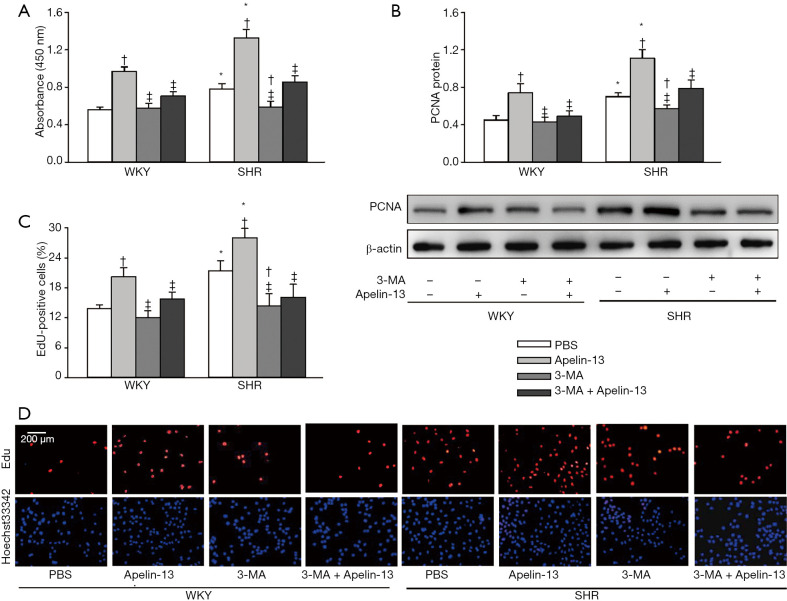
Figure 6.
Effects of autophagy inhibitor 3-MA on apelin-13-induced VSMC proliferation of WKY and SHR. (A) VSMC proliferation evaluated with CCK-8 kit. (B) VSMC proliferation evaluated with PCNA protein expression. (C) VSMC proliferation evaluated with the percentage of EdU-positive cells. (D) Representative images of EdU staining. Red, EdU-positive cells. Blue, Hoechst 33342 was used to label double-stranded DNA and thus to visualize nuclei. The measurements were carried out 24 h after PBS, apelin-13 (1 µM), or 3-MA (5 mM) treatment. Values are mean ± SE. *, P<0.05 vs. WKY; †, P<0.05 vs. PBS; ‡, P<0.05 vs. Apelin-13. n=6. 3-MA, 3-methyladenine.

