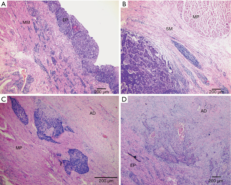
Figure 1.
Four categories of Japanese residual tumor pattern in esophageal wall tissue after NCRT using HE staining. (A) Type 1 (shallow remnant within EP to MM layers); (B) type 2 (central remnant within SM to MP layers); (C) type 3 (deep remnant within MP to AD layers); (D) type 4 (diffuse remnant from EP to AD layers). Scale bars in images, 200 µm. NCRT, neoadjuvant chemoradiotherapy; EP, epithelium; MM, muscularis mucosa; SM, submucosa; MP, muscularis propria; AD, adventitia.

