Fig. 2.
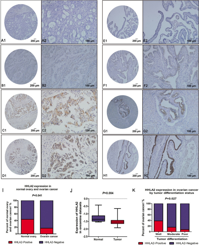
HHLA2 expression in ovarian cancer and normal ovary tissue. HHLA2 was stained with anti-HHLA2 antibody, and HHLA2-positive staining was detected in a membranous and cytoplasmic pattern in ovarian cancer and normal ovarian tissue epithelium. a–d represented epithelial ovarian cancer tissue obtained from 64 patients with ovarian cancer who had undergo surgical resection between 2007 and 2011. All patients were treatment-naïve before surgery. The level of HHLA2 expression was graded as intensity of IHC staining in ovarian cancer: A1-A2, absent; B1-B2, weak; C1-C2, moderate; D1-D2, strong. e–h The level of HHLA2 expression was graded as intensity of IHC staining in normal ovarian tissue epithelium: E1-E2, absent; F1-F2, weak; G1-G2, moderate; H1-H2, strong. Magnification: × 100 (A1–H1); × 200 (A2–H2). i The positive rate of HHLA2 expression was higher in normal ovary tissue than that in ovarian cancer. The red column represented the HHLA2 positive group, the blue column represented the HHLA2 negative group. The difference between them is significant.(p < 0.05). j Expression of HHLA2 was frequently decreased in 585 ovarian cancer tissues (Tumor) compared with 8 normal ovary tissue (Normal) samples in the oncomine dataset. k The proportion of HHLA2-positive patients was gradually increased with the improvement of tumour differentiation degree. HHLA2 tended to be expressed in well-differentiated ovarian tumour. The red column represented the HHLA2 positive group, the blue column represented the HHLA2 negative group. The difference between them is significant. (p < 0.05)

