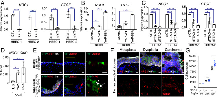
Fig. 6.
YAP/TAZ-TEAD directly regulate NRG1 expression. (A) Two HBEC lines, isolated from different patients (listed as HBEC-1 and HBEC-2), were transfected with control siRNA or siRNA targeting both YAP and TAZ. RNA was then isolated and analyzed by quantitative PCR for NRG1 and CTGF expression. (B) 16HBE cells were infected with lentivirus, transducing the expression of wild-type (WT) YAP or a nuclear-localized active YAP (5SA) and then analyzed by quantitative PCR for NRG1 and CTGF expression. (C) Two patient lines of HBECs were transfected with control siRNA or two different combinations of siRNA targeting all four TEAD family members, and isolated RNA from these cells was analyzed by quantitative PCR for NRG1 and CTGF expression. (D) YAP and TEAD ChIP was performed in human AALE bronchial epithelial cells and examined by quantitative PCR. (E) Airway epithelial tissues from control (LSL-YFP; K8CreERT2) mice and Crb3-cnull (Crb3-F/F; LSL-YFP; and K8CreERT2) mice isolated 21 d post-TMX treatment were examined by RNAscope for NRG1 mRNA and IF microscopy for YFP and Krt5. Elevated NRG1 expression was observed in YFP-marked Crb3-deleted cells (white arrows) and in neighboring nonYFP-marked Krt5-positive cells. (Scale bar, 20 μm.) (F) Human bronchial biopsies with the indicated pathology were examined by RNAscope for NRG1 mRNA levels and IF microscopy for Krt5 protein, revealing high NRG1 expression in precancerous Krt5-positive lesions. (G) Equal numbers of primary HBECs were treated with or without recombinant NRG1, and cell numbers were quantified at the indicated time points. Shown in the average ± SEM. Significance was determined with an unpaired t test. *P < 0.05, **P < 0.01, ***P < 0.001, and ****P < 0.0001.

