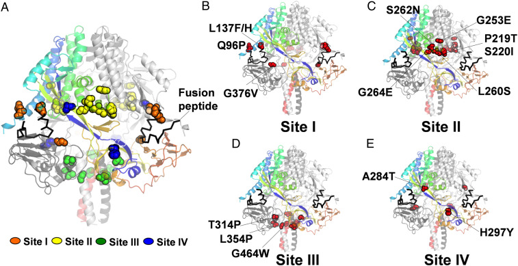
Fig. 5.
Structural mapping of hyperfusogenic F mutants. (A) The aa residues (spheres) whose substitutions confer hyperfusogenicity were mapped onto the trimeric prefusion MeV-F structure (Protein Data Bank: 5YXW) using Pymol. A ribbon model is shown in which each of the protomers is colored rainbow, dark gray, and light gray, respectively. (B–D) The majority of mutants discovered in this study, while novel, mapped to the three sites (I, II, and III) that were previously used to classify extant hyperfusogenic mutants by Hashiguchi et al. (E) Two mutants, A284T and H297Y, mapped to a structurally distinct beta sheet that connects the head and neck domains, which we term site IV. (A) In the aggregate model showing all the mutants, site I to IV mutants are represented by orange, yellow, green, and blue spheres, respectively.

