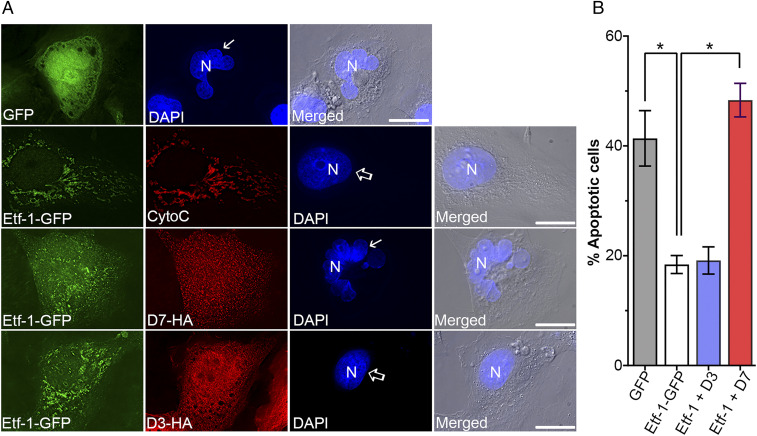
Fig. 6.
D7 abrogates Etf-1 inhibition of etoposide-induced apoptosis. RF/6A cells were transfected with GFP or Etf-1–GFP, or cotransfected with Etf-1–GFP and D7 or D3, followed by treatment with 100 µM etoposide at 24 hpt for 41 h. Cells were labeled with mouse monoclonal anti-cytochrome c (CytoC) and rabbit polyclonal anti–Etf-1, or mouse monoclonal anti-GFP and rabbit monoclonal anti-HA. (A) Images show the localization of Etf-1 and Nbs, and nuclear morphology was stained by DAPI (arrows, apoptotic nuclei; open arrows, nonapoptotic nuclei). Merged, the fluorescent image of DAPI channel merged with the DIC image. (Scale bars, 10μm.) (B) Quantification of apoptosis (nuclear fragmentation) in 100 cells expressing transfected genes from three independent experiments. Data are represented as the mean ± SD (n = 3). *P < 0.05, by one-way ANOVA.

