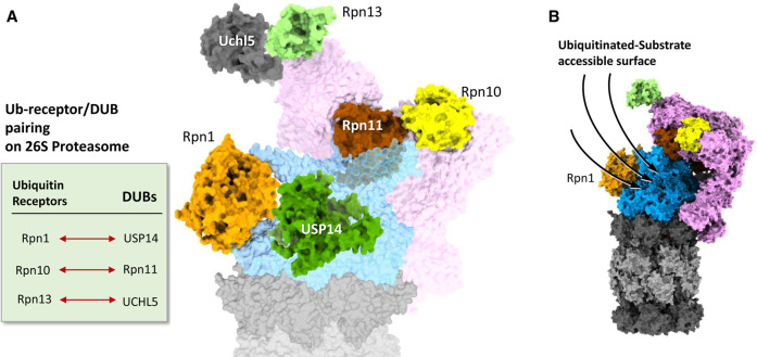
Figure 3. Features of 26S proteasomes adapted for substrate binding.
(A) Ubiquitin-receptor and deubiquitinase pairing on 26S proteasome. Ubiquitin receptors and deubiquitinases are positioned in pairs (Rpn1-USP14, Rpn10-Rpn11 and Rpn13-Uchl5). Models of 26S proteasome are by ChimeraX from PDB: 6j2x, Usp14 from 5gjq (Ubl-domain missing), and Uchl5 from 3ihr. Usp14 and Uchl5 models are simply placed over the proteasome model according to their corresponding positions. (B) A side view of a 26S proteasome showing the putative surface accessible for ubiquitinated substrate binding. Model of 26S proteasome is generated by ChimeraX using the PDB: 6j2x.

