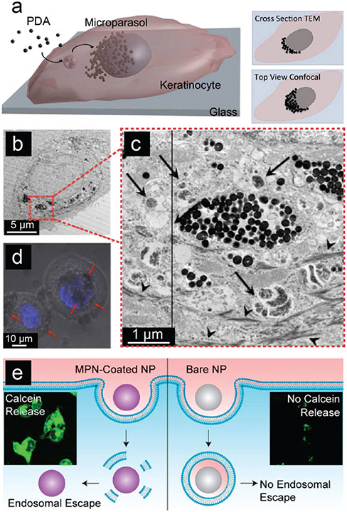
Fig. 18.
Cellular uptake study of phenolic nanoparticles. (a) Scheme for the uptake, transportation, and accumulation of PDA nanoparticles in cells. (b) TEM image, (c) enlarged TEM image, and (d) confocal laser scanning microscopy image of cells incubated with PDA nanoparticles for 3 days. (a–d) Reproduced with permission. Copyright 2017, American Chemical Society. (e) Scheme and fluorescence microscopy image of endosomal escape of bare and MPN-coated nanoparticles. Reproduced with permission. Copyright 2019, American Chemical Society.

