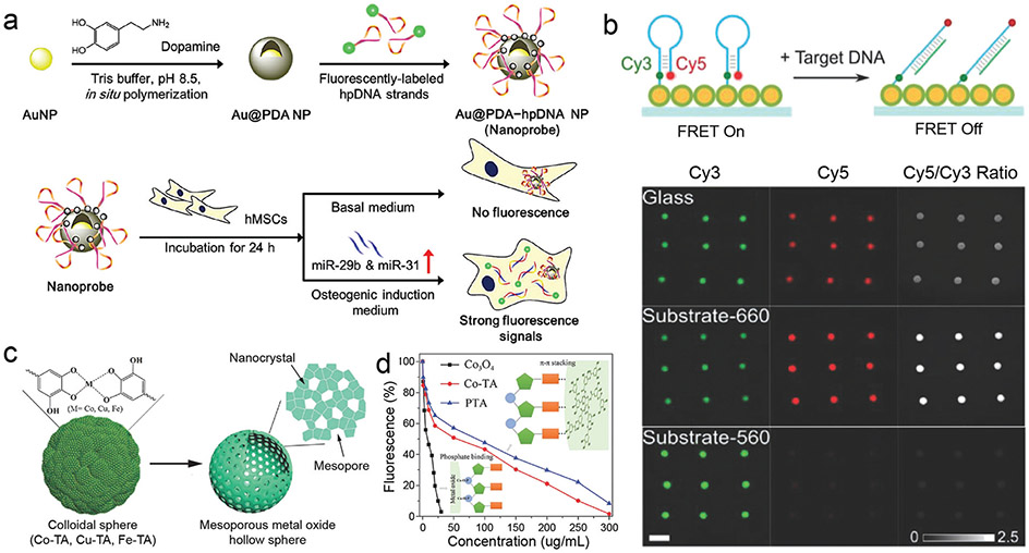
Fig. 21.
In vitro biosensing of biomacromolecules by using phenolic-based nanoparticles. (a) Schematic illustration of the synthesis of nanoprobes containing PDA-coated Au nanoparticles (Au@PDA NPs) and hairpin-DNA-based (pDNA), and their use for detecting miRNA targets in living hMSCs. Reproduced with permission. Copyright 2015, American Chemical Society. (b) Schematic illustration and fluorescence images of a DNA microarray of Cy3/Cy5 FRET on a plasmonic substrate. After binding a target DNA, the dye pair in the molecular beacon separated. Reproduced with permission. Copyright 2020, Wiley-VCH. (c) Schematic illustration of the synthesis of hollow MMOSs. (d) Fluorescence intensities of probe DNA depended on different concentrations of colloidal spheres. Insets are the schematic illustration of the interactions between probe and materials. (c and d) Reproduced with permission. Copyright 2018, Wiley-VCH.

