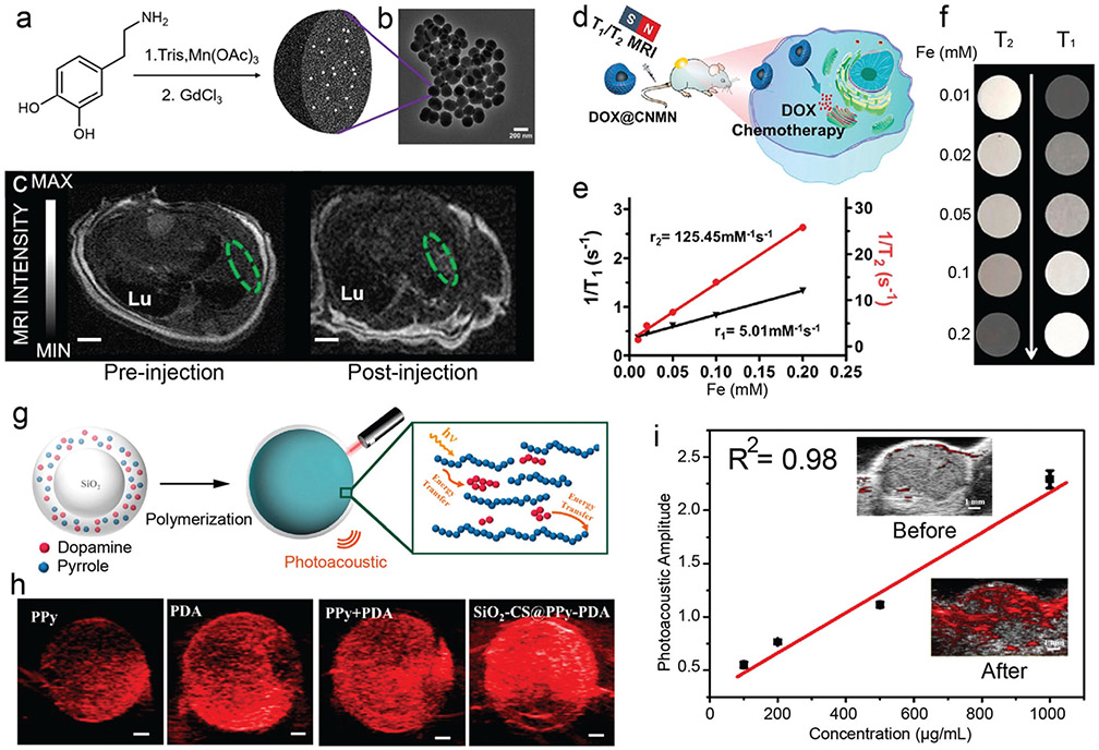
Fig. 23.
In vivo MRI and PAI performance of phenolic-based nanoparticles. (a) Synthesis and (b) TEM image of Gd3+-doped PDA nanoparticles. (c) Transverse MRI view of a mouse heart before and after injection of the MR CAs. Reproduced with permission. Copyright 2019, American Chemical Society. (d) Schematic illustration of T1/T2 MRI-guided cancer therapy by DOX-loaded TA-Fe(iii) coordination network (DOX@CNMN). (e) r1 and r2 relaxivities of CNMN as a function of the Fe molar concentration in the solution, and (f) the corresponding MR images of CNMN. (d–f) Reproduced with permission. Copyright 2020, American Chemical Society. (g) Schematic illustration of silica nanoparticles with PDA and PPy coating for PAI. (h) Photoacoustic images of suspensions of different particles and (i) photoacoustic amplitude of PPy–PDA-coated silica particles at different concentration. Inset: Photoacoustic images at 700 nm of the tumor before and after injection of the particles. (g–i) Reproduced with permission. Copyright 2018, American Chemical Society.

