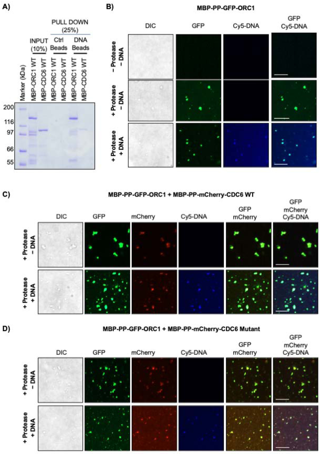
Figure 3. Phase separation of ORC1 and CDC6 proteins on DNA.

(A) MBP-ORC1 and MBP-CDC6 proteins were incubated with biotinylated dsDNA streptavidin beads in presence of 1mM ATP. Streptavidin beads alone served as control. (B) MBP-PP-GFP-ORC1 (4 μM) do not undergo a liquid-liquid phase separation without protease, but upon addition of protease in the absence or presence of 4 μM Cy5-dsDNA cleaved full length GFP-ORC1 forms liquid droplets. (C-D) MBP-PP-GFP-ORC1 (2 μM) protein was mixed with either 2 μM of MBP-PP-mCherry-CDC6 WT (C) or CDC6 Cy mutant protein (D) in the presence of protease without or with Cy5-dsDNA (4 μM). Merged images shows co-recruitment of cleaved GFP-ORC1 and mCherry-CDC6 (WT or mutant) with or without Cy5-dsDNA. See Figures S3C and S3D. In figures B-D, the images are representative of three independent experiments. The liquid droplets formed were captured after digestion with protease at 4°C for 16 hours. Scale bars 10 μm. PP; PreScission protease site.
