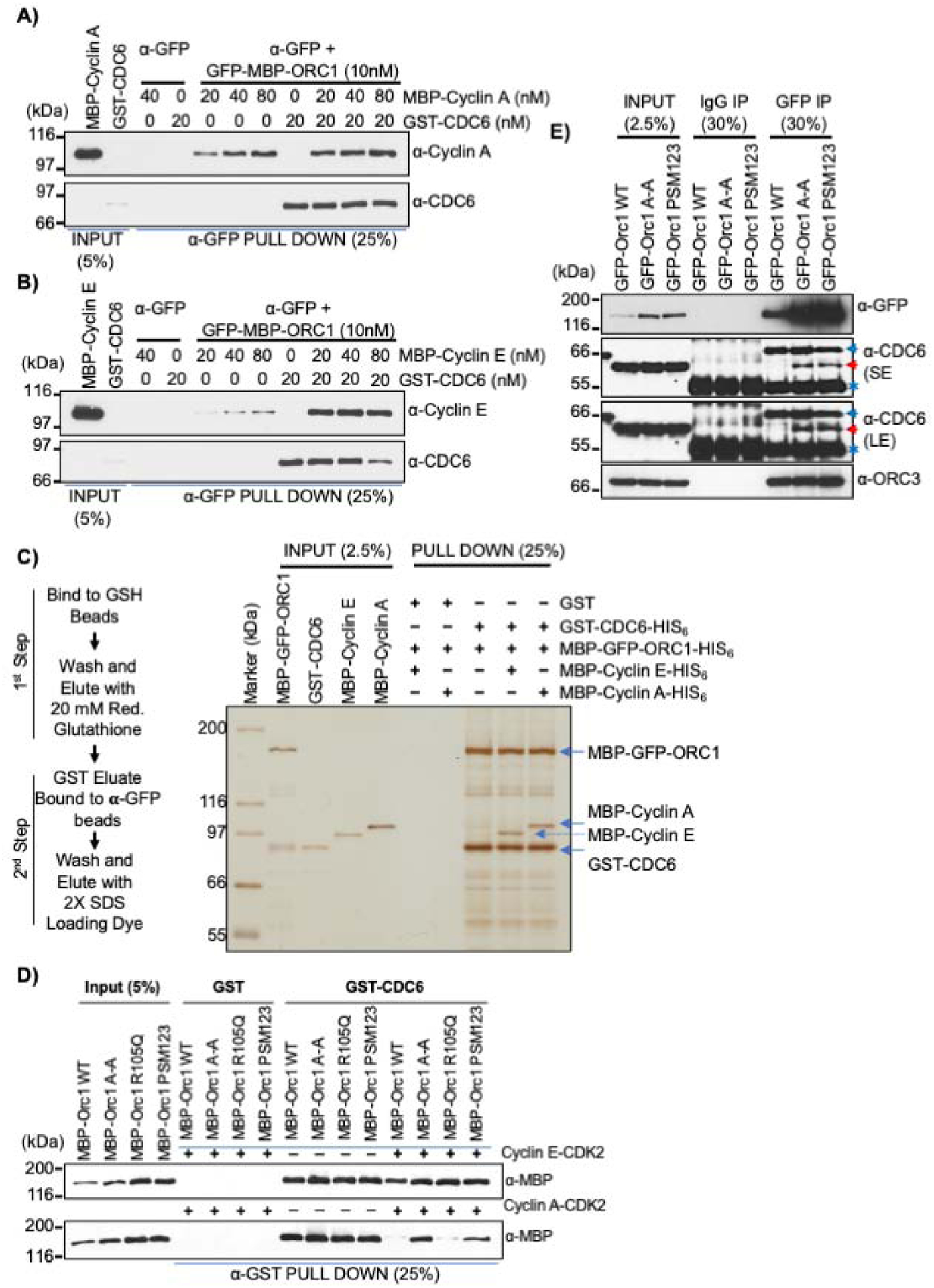
Figure 4. G1-S Cyclin-CDK kinases have differential effects on ORC1-CDC6 protein interactions.

(A-B) In a α-GFP antibody pull-down assay, a constant amount of recombinant GFP-MBP-ORC1 protein was incubated with GST-CDC6 alone or with increasing molar amounts of MBP-Cyclin A (A) or MBP-Cyclin E (B) and the levels of Cyclin or CDC6 co-precipitated were detected by immuno-blotting. See Figures S4C and S4D. (C) Schematic outline of a two-step affinity purification of protein complexes using purified recombinant proteins (Left panel). MBP-GFP-ORC1-HIS6 (50nM) and GST-CDC6-HIS6 (50nM) were incubated together or in combination with MBP-Cyclin E-HIS6 (50nM) or MBP-Cyclin A-HIS6 (50nM) and control GST (50nM) protein were incubated with either MBP-Cyclin E-HIS6 or MBP-Cyclin A-HIS6 and MBP-GFP-ORC1-HIS6 as indicated. After two step purification, the protein complexes were detected with silver stain (right panel). The reaction contained 1mM ATP. See Figure S4E and S4F. (D) In a GST pull-down assay, GST-CDC6 protein was incubated with MBP-ORC1 WT or its mutants in the presence or absence of Cyclin E-CDK2 (top panel) or Cyclin A-CDK2 (bottom panel) with 1mM ATP. The western blot is probed with anti-MBP antibody and GST protein served as negative control. (E) Mitotic cells from stable GFP-ORC1 WT or ORC1 mutant cell lines were collected after nocodazole treatment and cell lysates were used for immunoprecipitation with anti-GFP antibody. The samples were further immunoblotted with CDC6 antibody and ORC3 antibody. A red arrow indicates specific CDC6 band, while a blue arrow and an asterisk symbol indicate non-specific and cross-reactive heavy IgG bands, respectively. SE, short exposure and LE, long exposure.
