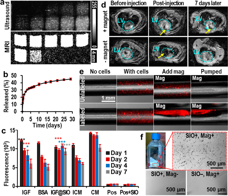
Figure 4.
Multiple functions of SIO. (a) Ultrasound and T2-weighted MR images of unlabeled (top row, the cell numbers are 0k, 10k, 50k, 150k, and 300k from left to right) and SIO-labeled hMSCs (bottom row, the cell numbers are 10k, 50k, 150k, 300k, and 600k from left to right). These images show the enhancement of both MRI and ultrasound signals of hMSCs after SIO labeling. MRI was done with a repetition time of 1400 ms and an echo time of 15 ms at 4.7 T. (b) Cumulative release profile of IGF from SIO. Error bars are standard deviations of triplicates. (c) In vitro survival of hMSCs treated with free IGF, free BSA, IGF-loaded SIO (IGF@SIO), incomplete media (ICM), and complete media (CM). No cells and nanoparticle-only groups are control groups with only incomplete media or media containing nanoparticles. Error bars are standard deviations of six replicates. The asterisks show the p value compared to incomplete media group; *p < 0.05, ***p < 0.0005. (d) MRI shows the long-term retention of SIO in the left ventricle wall only with the presence of a magnet. Azure dotted circles show the outlines of the left ventricles. Yellow arrows indicate the locations of SIO. (e) Overlay of fluorescence and microscope images show that SIO and magnets could improve the retention of hMSCs in laminar flow with a shear stress at 27 dyn/cm2. Both cells were stained with fluorescent quantum dots for visualization. (f) Suspended SIO-labeled hMSCs could be directed by an external magnet to the side wall of the flask. These attracted cells could adhere to and grow on the side wall. In contrast, no cells would grow on the same location without SIO labeling or without a magnet.

