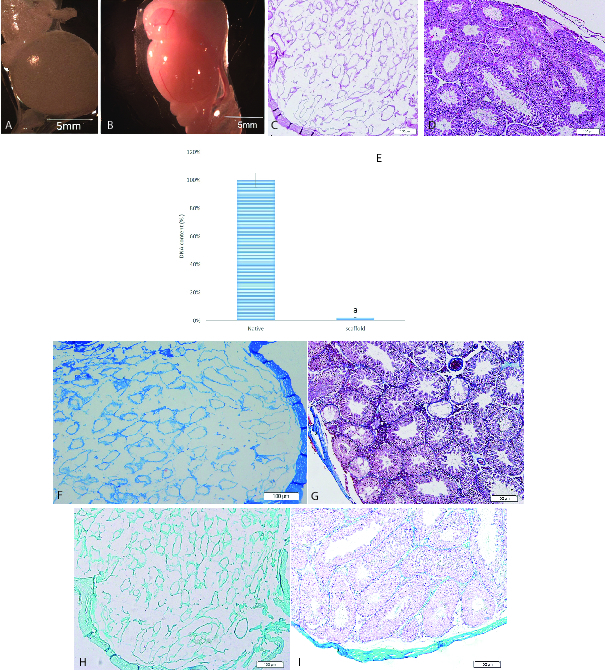
Figure 1.

Characterization of testicular scaffolds. Macroscopic images showed that scaffolds were completely translucent (A) while native testes were opaque (B). Histological comparison of scaffolds (C) and native testes (D) by H&E staining exhibited the elimination of the cells. Original magnification 100x. DNA quantification confirmed the removal of 98% of the DNA from the tissue (E). a indicated significant difference with native testis (p 0.05). Masson's trichrome staining showed collagen preservation in scaffolds (F) and native testes (G). Alcian blue staining confirmed GAGs retention in scaffolds (H) and native testes (I). Original magnification 100.
