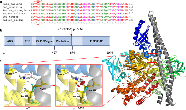
Fig. 3.
Structural analysis of PIK3CD. a Lysine 666 is highly conserved across multiple species. b PI3KCD encodes p110δ protein, which consists of an adaptor-binding domain (ABD), a Ras-binding domain (RBD), a C2 domain, an accessory domain (PIK helical) and a PI3K/PI4K catalytic domain. Mutation c.1997 T > C is located in the PIK helical domain. c Three-dimensional structure of p110δ shown in colour. After the alteration of L666P, repulsion will occur between Pro 666 and Arg 663 as well as Pro 666 and Leu 662, resulting in the instability of protein structure

